 Gamma Knife or gamma-ray bistoury is a revolutionary instrument
which allows treating vascular malformations, such as tumors, and some
functional brain pathologies, without needing to open the brain.
Gamma Knife or gamma-ray bistoury is a revolutionary instrument
which allows treating vascular malformations, such as tumors, and some
functional brain pathologies, without needing to open the brain.
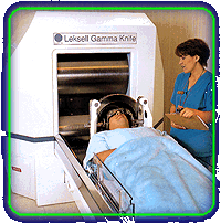
Many of these lesions which were previously regarded as untreatable, can now be treated with Gamma Knife. The equipment uses 201 cobalt-60 concentrated rays which are focused with an extreme precision on the target to be treated, in order to destroy it without lesioning the neighboring brain tissue. Every penetrating ray is silent, that means, that it does not harm by itself. It is the convergence point of the 201 rays where the maximal energy concentration is given, and the effect on the target is exerted. The treatment is performed by a multidisciplinary group of professionals, including neurosurgeons, a radio-oncologist, a radiation physicist, technicians, and specialized nurses whose objective is precision with an optimal quality control when attending the patient. How does Gamma Knife work? At Hospital San Javier, we understand there is fear due to the procedures, and simply due to the fact of knowing that the diagnosis of a tumor or a cerebral lesion was given. That is why we explain step by step the procedure to carry out a treatment with Gamma Knife. Step 1: Adjustment of stereotactic frame By means of small screws, a metallic frame, the so-called stereotactic frame, helps us defining the target to be treated, and preventing that the head moves during the treatment procedure. A small dose of anesthesia is used to diminish discomfort during the adjustment of the stereotactic frame to your head. This frame is used during the treatment planning and it determines the precise coordinates of the zone or target in the head to be treated. Step 2: Images After adjusting the frame, the following step is performing the image studies of your brain, in order to locate the abnormality. Image studies may be performed by means of magnetic resonance, computerized tomography, or angiography, or a combination of these studies, depending on the kind of alteration to be trated. Step 3: Computerized treatment planning Once the brain image studies have been completed, these are transferred to the computerized treatment planning system. The Gamma-Knife specialists use a computer to determine the exact coordinates of the brain alteration (or tissue), and to calculate how many radiation shots will be necessary for the treatment. This will help covering the size of the abnormality as tight as possible with an proper radiation dose. Step 4: Treatment Now you are ready for the treatment. During the procedure, you will be in a constant auditive and visual communication with the specialists and the physicians treating you. You will be placed on the Gamma-Knife bed, and the stereotactic frame, which is fixed to your head, will be adapted to a great metallic helmet, containing the collimators. Having the shape of a great hemisphere, the collimator helmet has many holes receiving and guiding the gamma radiation to the area of the brain which has been selected for the treatment. Once you have been correctly positioned, the Gamma Knife will administer 201 rays to the tissue or abnormality. Each ray by itself is not powerful enough to cause any harm, but at the place where the 201 rays join (the area of abnormality or target point) the radiation is powerful enough to treat the zone. Step 5: Recovery and leaving the hospital Once the treatment has been completed, the stereotactic frame is removed, and bandages are placed where the screws were fixed. You stay at the hospital one night for observation. The bandages are removed the following day. The small places where the screws were fixed heal very quickly, and they can be washed with soap and water 48 hours after the treatment with Gamma Knife. If there is numbness or pain at the places where the screws were fixed, these disturbances disappear after a couple of weeks. The follow-up given to the patients treated with Gamma Knife include visits to the referring physician, and the Gamma-Knife team physicians. They will watch over your evolution by means of visits and image studies such as the magnetic resonance of the cranium, as well as other studies which are considered pertinent.
|
. |
A New Concept of Surgery without a Bistoury |
|
[Support of HSJ]--[revious Considerations]--[FAQ's]--[Do you need more Information?]
|
||